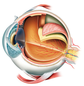
Ocular muscles of the human eye vision system.
This short video demonstrates how the eye muscles work together to move the eye. Get my new (May 2013) interactive book on your iPad, http://itun.es/i6xT3Yf …
Ocular muscles and the actions of the eyes
Ocular muscles of the human eye vision system are fascinating to study, here is a short animated video with the actions of the ocular muscles moving the eyes. Watch this short video prepared and presented to you on the YouTube channel.

Actions of the eyes by the ocular muscles
There are four muscles per eye that move the eyes up and down, and side to side.
Names of three muscles are:
- Medial rectus.
- Inferior Oblique
- Superior Oblique
- Lateral Rectus
- Superior Rectus
- Inferior Rectus.
Ocular muscles do need a special attention to make sure they line up the eyes accurately, if one eye is just slightly out of line the result will be a blurred vision or double vision. The ocular muscles need to line up both eye precisely on the focused object so that the visible object is seen as one singular object. Learn more on how to take care of your eye vision for life, visit this link now.
Ocular muscles contraction and relaxation
Four of the ocular muscles control the movement of the eye in the four cardinal directions: up, down, left and right. Ocular muscles contract and the opposite side muscles relax at the same time, enabling a synchronization of the eye movement. See video for clear presentation of the eye movements.
Essential nutrients for the eye vision
Essential nutrients are necessary for a healthy eye vision, similar all other biological systems of the human body they need a constant supply of life-giving nutrients. Essential nutrients for the eye vision are also manufactured into a supplement form. health eating habits ensure that the body receives all of the necessary nutrients that all of the eleven biological organ systems need.

really helpful in my study..thx
When the eyes are adducted, the inferior oblique muscle is responsible for
elevation? I don’t quite understand the intuition behind this anatomy..
Good video but you need to hydrate yourself before speaking on videos dude.
Squelchy sounds are offputting!
TAHNKS.
This is amazing work. The animations are perfect, your explanation is
clear, eloquent and succinct. Massive respect to you and your work. Thank
you.
Thxs for good work. It’s really amazing :D
Brilliant. Amazing.
Very nice muvie recommended by http://www.okulista.wroclaw.pl
Sir- Im a little confused with the last diagram. From my understanding, the
inferior oblique muscle kicks in during the abducted phase of the eye. In
other words, when the lateral rectus abducts the eye, elevation occurs
because the inferior oblique contracts and not the superior rectus.
However, this diagram shows that when the medial rectus contracts
(adduction) the inferior oblique elevates the eye. Could you perhaps
clarify this. Thanks.
woweee, great video!
Your diagram is misleading, in that the SO and IO are shown medially
(between the two eyes) – SO and IO also partially abduct the eye, in
isolation and in clinical testing are always drawn outwards for the
classical ‘H’ testing of EOM. Other than that, cool video.
Really helpful, explained my lecture notes perfectly. Diagram at the end is
particularly useful – thanks you!
Models done in blender.
thank u v much…really helps alot…thks…^.^
I believe you have the contractions/relaxations backwards at 1:30
The last picture is 100% right, but I need a way to better explain it. I’m
discussing the labelled diagram at the end. This shows you how to remember
the MAIN ACTIONS of the muscles. Medial and lateral rectus pull the eye in
and out, so there is no confusion there. With the eye already abducted,
sup. rectus elevates, inf. rectus depresses the eye. With the eye adducted,
inferior oblique elevates and sup oblique depresses. Read the transcript,
or read my ibook…(will be in iBookstore soon)
@poobah42o no, your statement disagrees with what the video says. stapsell
is correct that the SR and IR elevate and depress the eye, respectively,
when the eye is ABducted, not ADdcuted (1:58). he is using a right eye in
his example.
Excellent and educational!
Because people are idiots
thank u so much, i’mma give u head now!
oh okay, thank you
I’ve spent so long trying to find some logic to the EOM rather than just
memorizing which muscle does what… thank you so much!